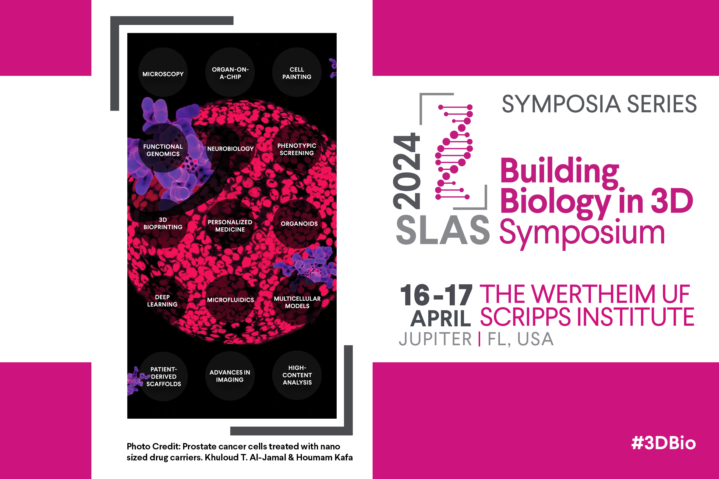
April 16-17, 2024
The Herbert Wertheim UF Scripps Institute for Biomedical Innovation & Technology
Jupiter, FL, USA


April 16-17, 2024
The Herbert Wertheim UF Scripps Institute for Biomedical Innovation & Technology
Jupiter, FL, USA
This session will explore the development and application of the latest innovations in imaging hardware and software for extracting knowledge from 3D model systems. We will discuss best practice tools and techniques for imaging in 3D including:
We will present case studies demonstrating tangible examples of when 3D imaging is required and provides an added benefit over traditional 2D assays. Finally, we will discuss unresolved challenges of 3D imaging and analysis and where concerted multidisciplinary research effort is required to advance the field.
Marc Bickle, Ph.D. (Roche)
Bickle obtained his Ph.D. at the Biozentrum in Basel, Switzerland, studying the immunosuppressive drug Rapamycin. He then went to the Laboratory of Molecular Biology in Cambridge, UK, to study the genetics of behavior in C. elegans. Afterward, he participated in the creation of Aptanomics, a drug discovery Biotech in Lyon, France, developing yeast 2 hybrid screens to select a collection of peptide aptamers. After this period in biotechnology, he headed the High-Content Screening facility at the Max Planck Institute of Molecular Cell Biology and Genetic (MPI-CBG) in Dresden, Germany, performing functional genomic and chemical screens using automated microscopy. Since 2021, Marc has been leading the Organoid Phenotyping platform at the Institute for Human Biology at pRed, Roche in Basel, Switzerland.
Kimberly Homan, M.S., Ph.D. (Genentech)
Leveraging 3D Imaging of Organoids for Basic Biology and Drug Screening Applications
Wei Ouyang, Ph.D. (KTH Royal Institute of Technology)
Building Web & Cloud-Based Platforms for AI-powered BioImage Analysis
Wei Ouyang is an assistant professor in the Department of Applied Physics at KTH Royal Institute of Technology (Stockholm, Sweden). He is currently leading a research group, the AICell Lab, which is funded by the Data-Driven Life Science fellow program. The group focuses on building AI systems for cell and molecular biology.
Dr. Ouyang obtained his PhD in computational image analysis at Institut Pasteur, Paris, where he mainly focused on applying deep learning for super-resolution microscopy. During this time, he developed a deep learning method called ANNA-PALM which massively accelerates super-resolution localization microscopy by 100x. He spent 4 years at Emma Lundberg’s group as a postdoctoral researcher.
Emil Rozbicki, M.S., Ph.D., M.B.A. (Glencoe Software)
Strategies for Scaling Image Data Analysis: Leveraging OME-Zarr and OMERO for Bioimage Data Access